
As medical professionals we understand the intricate and nuanced process of treating a patient from start to finish. From our first encounter taking a detailed history, to completing a physical examination, to ordering appropriate tests, to making a diagnosis and treatment plan, we make decisions based on the best information available to us at that time.
In most cases, during an initial exam or interaction we evaluate the patient based on what can be seen externally, heard with a stethoscope, and felt by palpating the body.
However, imaging technology has recently advanced to the point where we are gaining the ability to ascertain more information during the physical exam—information previously unavailable without additional testing or a referral.
Innovations in ultrasound
For years, increasingly portable ultrasound technology has made the power of imaging more broadly available in a variety of medical settings. While these ultrasound machines are helpful, they have not been readily accessible to physicians. However, new innovations in the field of ultrasound are adding a dimension to the physical exam that could potentially aid in improving and enhancing the care delivery process—the ability to see inside of a patient during that initial exam.
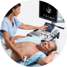
Vscan, a pocket-sized ultrasound developed by GE Healthcare for use by physicians, has brought ultrasound technology to about the size of a cell phone or PDA. Vscan provides physicians with the ability to take high-quality internal images of a patient’s body directly at the point of care in the exam room.
Vscan can be used to visualize and assess the major vessels and organs such as the aorta, heart and lung, liver, spleen, kidneys and the chest, abdomen, and pelvis for free fluid. Given its size, Vscan can easily be carried from exam to exam, room to room, and used on multiple patients (with appropriate infection control measures in place, of course).
Enhancing our tried-and-true methods
While the traditional examination methods of observing, listening, and feeling remain crucially important to the way in which we examine, diagnose, and determine treatment options for our patients, this new tool could potentially redefine the way we conduct exams. The capability to immediately obtain internal information on a patient during the exam permits physicians to add an extra layer of information as they assess a patient. For example, for an abdominal injury, an early focused assessment with sonography in trauma (FAST) examination provides crucial information on whether there is free blood
in the abdominal cavity. Immediate clinician-performed point-of-care testing can help ascertain if a traumatized patient is appropriate for further imaging or rapid transfer to the operating room.
Even during a non-emergency routine exam, such as a surgical consultation for abdominal pain or a yearly exam conducted by a primary care physician, the ability to obtain an immediate image could help to facilitate diagnostic decisions and management plans.
A portable ultrasound is also useful for frequent monitoring of the patient before transfer to definitive surgical or medical care. One can be looking for signs of change of the initial FAST imaging without having to repeat more expensive procedures before transfer or holding in the emergency department, providing some degree of comfort that things really are stable.
Of course, this is not to say that a pocket-sized ultrasound replaces ad-vanced, clinical diagnostic imaging performed and interpreted by expert technicians and radiologists, or that it alone can diagnose a condition—this is, of course, always up to the discretion and expertise of the physician. However, with proper training and experience, it may guide decision making around the need for and timeliness of further imaging.
Scenarios of use
During the winter of 2010, we had the privilege of serving as, respectively, the Whistler Polyclinic manager for the Vancouver 2010 Olympic and Paralympic Winter Games and as chief medical officer.
At the Whistler athletes’ Village, we kept the Vscan on hand for the use of our emergency physicians and surgeons who had experience and credentialing with clinician-performed ultrasonography in their regular practices. The pocket-sized ultrasound was used routinely for scanning appropriate patients seen in the Polyclinic, including those with multi-trauma or single-system complaints of abdominal or chest pain.
The speed of access, quality imaging, and portability of the instrument made it well suited for initial assessments in a very small trauma and resuscitation bay within the Mobile medical unit, a trailer adjacent to and part of our temporary tent facility that made up the Polyclinic. Despite having 24/7 access to innovative equipment and outstanding radiology technicians and radiologists, a preliminary study with the handheld device was often completed before our larger imaging machines were ready for use.
For example, in this typical (fictitious) clinical scenario, a 25-year-old patient sustained significant blunt force trauma from a fall during a high-speed alpine skiing event. The patient had an obvious extremity fracture and was complaining of abdominal and chest pain. Following rapid scene assessment, treatment, immobilization, and evacuation, the patient arrived in the Polyclinic resuscitation bay within minutes of the incident.
Using advanced trauma life support principles, the trauma team performed the ABCs of a primary survey moving on to a secondary head-to-toe survey.
Notwithstanding the patient’s initial stable vital signs, the trauma team leader wanted to determine if there was objective evidence of intra-abdominal bleeding while concurrently preparing the patient for further imaging and transfer to definitive care. A bedside physician-performed FAST with Vscan was completed in minutes, giving the trauma team leader a degree of comfort before allowing the patient to be moved to the CT scanner for further imaging and, ultimately, a helicopter evacuation to the Vancouver General Hospital for definitive care.
In another fictitious example, a male skeleton athlete was brought into the mobile medical unit from the sliding centre after hitting a wall very hard on his left chest and upper abdomen. With Vscan, fluid was visible around the spleen. As the patient was hemodynamically stable, he was taken to the CT scanner, where an abdominal CT confirmed a splenic laceration with no active bleeding. The patient was then prepared for helicopter transfer to the Vancouver General Hospital’s trauma unit for definitive care.
Beyond patient care
An added benefit to point-of-care imaging with portable ultrasound is that it can be used as a teaching tool. The medical students and surgical residents we teach at the bedside are quick to understand the benefits of this technology. It provides additional information, facilitating their understanding of an individual patient case, and allows them to better link their knowledge of anatomical structure and function with the pathological or normal findings they are seeing with the device.
Conclusion
Although traditional methods of examination are tried and true, innovations in ultrasound are enhancing the ways in which we evaluate and eventually treat patients.
We are looking forward to seeing the capabilities of the next generation of hand-held ultrasound devices and using them to help optimize patient care.
Competing interests
Vancouver General Hospital and Dr Brown have had access to the Vscan on consignment from GE Healthcare as a part of the Vancouver 2010 Winter Games Legacy project. Dr Brown has not received any financial compensation from GE Healthcare and does not have any corporate interests in the company. Dr Taunton was invited to speak at the Canadian Health Informatic Awards with GE as the winner of the Project of the Year with 2010 Olympic Winter and Paralympic Games.
Dr Brown is a commander in the Canadian Forces Reserve, a general and trauma surgeon, and clinical professor at the University of British Columbia. He was the manager of the Whistler Polyclinic for the Vancouver 2010 Olympic and Paralympic Winter Games. Dr Taunton is a professor in the Division of Sports Medicine, Faculty of Medicine at the University of British Columbia and sport medicine physician at the Allan McGavin Sports Medicine Centre. He was the chief medical officer for the Vancouver 2010 Olympic and Paralympic Winter Games.
Source URL: http://bcmj.org/articles/using-pocket-sized-ultrasound-tools-patient-care
Links:
[1] http://bcmj.org/author/ross-brown-omm-cd-bsc-ma-md-frcsc-facs
[2] http://bcmj.org/author/jack-taunton-msc-md